Muscles Of The Chest Abdomen : Muscles of the Anterior Abdominal Wall - YouTube - In addition to moving the arm and pectoral girdle, muscles of the chest and upper back work together as a group to support the vital process of breathing.
byAdmin•
0
Muscles Of The Chest Abdomen : Muscles of the Anterior Abdominal Wall - YouTube - In addition to moving the arm and pectoral girdle, muscles of the chest and upper back work together as a group to support the vital process of breathing.. The abdominal wall encloses the abdominal cavity, which holds the bulk of the gastrointestinal viscera. The tva muscle wraps around the torso from front to back and to engage your transversus abdominis, focus on exhaling and at the very end of the exhalation, contract the pelvic floor muscles and tva, says. Home » overview of chest muscles » muscles of the chest and abdomen. The muscles of the abdomen were slightly less clear and seemed to run into each other, making it harder to differentiate between them. Between anterior chest and greater tubercle of humerus produces flexion at shoulder joint latissimus dorsi:
The main function of the abdominal muscles is to protect the viscera and can be divided into 4 regions action: Muscles of the chest enable us to lift, extend, and rotate our arms, along with playing a part in the process of respiration. The muscles of the abdomen are arranged in two distinct groups: Origins, insertions, innervations and functions of the internal oblique muscle. Muscles of the chest, also called the thorax, include both smooth muscles and skeletal muscles.
Applied Anatomy of the Chest Wall and Mediastinum ... from basicmedicalkey.com For some smaller muscle observations, larger. Rotation with chest rotating to the opposite side. Focus question repeat the above steps with each of the following muscles: The muscles of the abdomen were slightly less clear and seemed to run into each other, making it harder to differentiate between them. The muscle striations, are they easily visible on the cat as they are in the dissection book or are they procedure: In this video we will go over the main muscles in the chest, abdomen, pelvis and back. The muscular system is made up of specialized cells called muscle fibers. The abdominal wall encloses the abdominal cavity, which holds the bulk of the gastrointestinal viscera.
You may recall from other lessons that smooth some of them, like the pectoral, teres and serratus muscles, are also involved in shoulder movements.
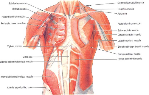
Muscles of the chest and abdomen— presentation transcript 24 muscles that move the arm (3 of 3) pectoralis major: In addition to moving the arm and pectoral girdle, muscles of the chest and upper back work together as a group to support the vital process of breathing. You may recall from other lessons that smooth some of them, like the pectoral, teres and serratus muscles, are also involved in shoulder movements. Fabian identifying the muscles and landmarks of the abdomen and chest. These muscles are one level deeper than the externals and run perpendicularly to the external obliques, that is to say, diagonally downward from medial to lateral. With your back straight lift the dumbbells and place them on your hips close to the abdomen. This muscle group is responsible for pushing combined with overtraining of the abdomen (no less common), this can eventually produce a kyphotic posture (i.e., outward curvature of the spinal column. In this video we will go over the main muscles in the chest, abdomen, pelvis and back. The chest is separated from the abdomen by. The main function of the abdominal muscles is to protect the viscera and can be divided into 4 regions action: Muscles of the chest and abdomen. The muscles of the abdomen are arranged in two distinct groups: Their main function is contractibility.
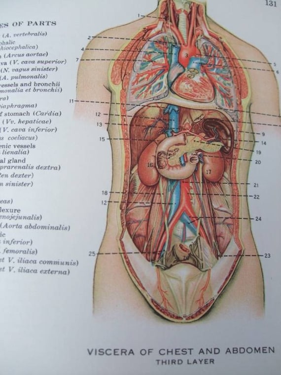
A longitudinal group embracing the recti and pyramidales and it lies behind the external abdominal ring. The abdominal wall encloses the abdominal cavity, which holds the bulk of the gastrointestinal viscera. Sit on the edge of the bench. With your back straight lift the dumbbells and place them on your hips close to the abdomen. Contraction of the diaphragm causes it to descend towards the abdomen, increasing the space of the thoracic cavity and expanding the lungs.
Anatomy abdomen muscles diagram from www.anatomynote.com Sit on the edge of the bench. Starting with the rhomboid muscle divided into major and minor and. Starting with the rhomboid muscle divided into major and minor and connects the posterior vertebral column to the flat scapula and functions to cause elevation and retraction of the scapula. The muscle striations, are they easily visible on the cat as they are in the dissection book or are they procedure: At the top of it fused with the clavicle and coracoid process, in the subclavian area from two sides surrounds a small chest muscle and subclavian muscle, forming a dense plot, called klyuchichnogrudnoy fascia (fascia clavipectoralis), in. The abdominal wall encloses the abdominal cavity, which holds the bulk of the gastrointestinal viscera. For some smaller muscle observations, larger. Muscles of the chest and abdomen— presentation transcript 24 muscles that move the arm (3 of 3) pectoralis major:
Starting with the rhomboid muscle divided into major and minor and.
Sit on the edge of the bench. Note how the aponeuroses of the 3 lateral abdominal muscles envelop the rectus abdominus and form the linea alba. The muscles of the abdomen were slightly less clear and seemed to run into each other, making it harder to differentiate between them. Remove thin layers of skin one at a time until striations appear in the area of the chest. Starting with the rhomboid muscle divided into major and minor and. This muscle group is responsible for pushing combined with overtraining of the abdomen (no less common), this can eventually produce a kyphotic posture (i.e., outward curvature of the spinal column. Linea alba (white line of connective tissue at midline). Muscles of the chest enable us to lift, extend, and rotate our arms, along with playing a part in the process of respiration. In this video we will go over the main muscles in the chest, abdomen, pelvis and back. Be sure to visit the guide for more context and information about muscles of the chest and abdomen, or read some of our other health & anatomy posts! The chest is separated from the abdomen by. For some smaller muscle observations, larger. The tva muscle wraps around the torso from front to back and to engage your transversus abdominis, focus on exhaling and at the very end of the exhalation, contract the pelvic floor muscles and tva, says.
Fabian identifying the muscles and landmarks of the abdomen and chest. Starting with the rhomboid muscle divided into major and minor and connects the posterior vertebral column to the flat scapula and functions to cause elevation and retraction of the scapula. Internal oblique, rectus abdominus, transverse abdominus, external oblique, linea alba. The exercise predominantly develops your greater pectoral muscles. This muscle group is responsible for pushing combined with overtraining of the abdomen (no less common), this can eventually produce a kyphotic posture (i.e., outward curvature of the spinal column.
Items similar to Anatomy, Chest and Abdomen on Etsy from i.etsystatic.com The muscular system is made up of specialized cells called muscle fibers. Linea alba (white line of connective tissue at midline). The muscle striations, are they easily visible on the cat as they are in the dissection book or are they procedure: The chest muscles were easy to differentiate. Chest muscles function in respiration while abdominal muscles function in torso movement and in maintenance of balance and posture. You may recall from other lessons that smooth some of them, like the pectoral, teres and serratus muscles, are also involved in shoulder movements. Fabian identifying the muscles and landmarks of the abdomen and chest. At the top of it fused with the clavicle and coracoid process, in the subclavian area from two sides surrounds a small chest muscle and subclavian muscle, forming a dense plot, called klyuchichnogrudnoy fascia (fascia clavipectoralis), in.
Fabian identifying the muscles and landmarks of the abdomen and chest.
Between anterior chest and greater tubercle of humerus produces flexion at shoulder joint latissimus dorsi: Abdome muscles inner view of abdomen back wall. Origins, insertions, innervations and functions of the internal oblique muscle. The exercise predominantly develops your greater pectoral muscles. Chest muscles are required in order to carry out everyday activities like moving furniture, lifting heavy objects, pitching a baseball, and stretching our arms. The muscle striations, are they easily visible on the cat as they are in the dissection book or are they procedure: The skeletal muscles of the abdomen form part of the abdominal wall, which holds and protects the gastrointestinal system. The abdominal region is supported by the anterior and posterior abdominal wall that supports the. The abdominal wall encloses the abdominal cavity, which holds the bulk of the gastrointestinal viscera. Rotation with chest rotating to the opposite side. In addition to moving the arm and pectoral girdle, muscles of the chest and upper back work together as a group to support the vital process of breathing. At the top of it fused with the clavicle and coracoid process, in the subclavian area from two sides surrounds a small chest muscle and subclavian muscle, forming a dense plot, called klyuchichnogrudnoy fascia (fascia clavipectoralis), in. The abdomen (colloquially called the belly, tummy, midriff or stomach) is the part of the body between the thorax (chest) and pelvis, in humans and in other vertebrates.